Study Update: Use of advanced magnetic resonance imaging to diagnose brachial plexus injuries and inform surgical intervention
The brachial plexus is a network of nerves which extend from the spinal cord to both hands and allow movement and feeling. A brachial plexus injury occurs when these nerves are stretched, compressed, or in the most serious cases, ripped apart or torn away from the spinal cord. 1% of patients involved in major trauma will have a brachial plexus injury, but current diagnostic techniques to aid clinical decision-making are not sufficiently accurate.

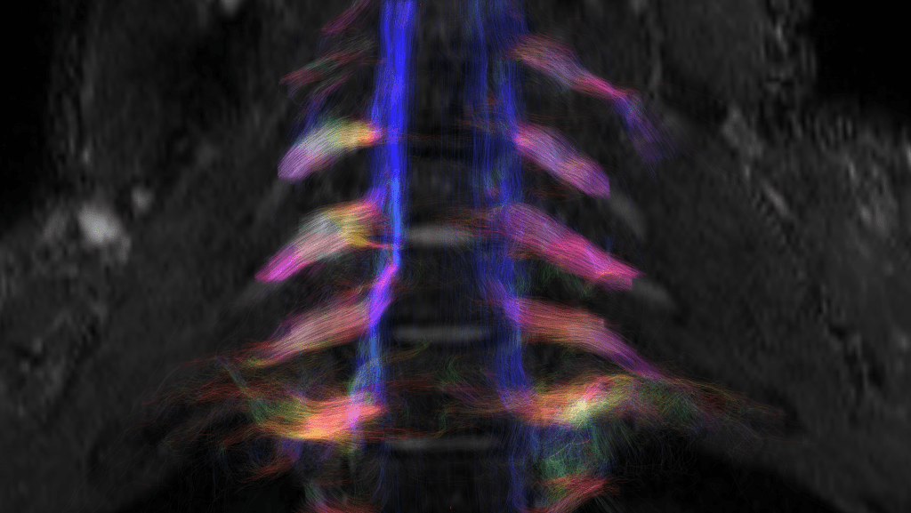
An advanced MRI of the brachial plexus
Magnetic resonance imaging (MRI) is currently used to help diagnose brachial plexus injuries, but traditional imaging protocols do not deliver the depth of information required by clinicians due to the complex ramified structure and the location of the brachial plexus. There is a need for advanced imaging techniques to show a detailed picture of the brachial plexus, especially whether there is continuity of the nerve to contribute valuable information on brachial plexus injuries to inform diagnosis and surgical intervention.
The Herston Imaging Research Facility (HIRF) secured a loan from Siemens Healthcare for an advanced MRI protocol, ‘SMS-RESOLVE diffusion imaging sequence’. A grant from HIRF allowed the advanced MRI to be performed and refined on ten healthy control participants and then eight patients who had sustained a brachial plexus injury underwent the study MRI. The Jamieson Trauma Institute (JTI) co-contributed to this study by providing research coordination for the duration of the project.
The study’s hypothesis is that the information provided by the new protocol will significantly aid with clinical decision-making and has four aims:
Aim 1. To compare the information obtained on the status of the brachial plexus against conventional diffusion and structural acquisitions.
Aim 2. To determine the value of the imaging information provided by an advanced MRI imaging protocol in showing continuity of the nerve and signs of recovery within the nerve.
Aim 3. To determine how the imaging information provided by this imaging protocol relates to study specific neurophysiology and clinical findings.
Aim 4. To correlate the pattern of injury shown on the HIRF MRI with neurophysiology measurements
During 2022-2023 10 patients with this injury were recruited on to this study, eight of whom successfully had an advanced MRI at HIRF. Information from their clinical examination (including neurophysiology reports) have been collated and discussed in relation to their imaging results. The study’s endpoints are to assess the ability of advanced MRI to deliver information about nerve continuity in brachial plexus injuries and to provide information that relates to clinical and neurophysiology findings. The study findings are currently being analysed as part of a Master’s program at the Centre for Advanced Imaging at the University of Queensland, with reporting expected by early 2025. It is anticipated that this study will provide an initial qualitative assessment of the imaging protocol and support a large quantitative study to deliver evidence of the benefits of advanced imaging in brachial plexus injury diagnosis.
