HIRF helps researchers see disease
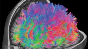
MR diffusion tractography showing the white matter tracts within the brain. These images are used to work out the connections between different parts of the brain and can be used clinically to guide neurosurgery. They are frequently used in brain imaging research at HIRF.
Director Paul Thomas says the facility is an extension of the hospital, with nuclear medicine and radiation technicians all hospital staff who rotate through HIRF. The facility is an alliance between Metro North Hospital and Health Service, the University of Queensland, QIMR Berghofer, and Queensland University of Technology all equal partners.
HIRF has three scanners for research. The advanced technology in the Magnetom Prisma 3T magnetic resonance imaging (MRI) machine was developed
specifically for the Human Connectome Project to map and understand the brain. HIRF has one of the few Prisma MRIs in Australia.
“This advanced MR technology allows higher quality MR scans and shorter scan times,” Paul says.
HIRF also has two scanners for positron emission tomography (PET) imaging. One of these is a combined PET and MR scanner, one of only a few in Australia.
“The PET-MR combination is a technological tour de force. It allows PET and MR scans to be performed at the same time, opening up new opportunities for research,” Paul says.
Dedicated to research, the high tech HIRF facility provides researchers access to equipment that in a hospital setting would be tied up with acute patients. Paul says although activity is still building, the relatively new facility has made a significant difference for Queensland researchers, with 40 projects already underway or completed.
Researchers come from each of the HIRF partners, external institutions and commercial entities. One research project made possible by access to HIRF’s technology is a longitudinal study of 450 people to identify MR, PET and other markers of Alzheimer’s disease that could help clinicians identify the disease as early as 10-20 years before conventional diagnosis.
Other researchers are looking at imaging different cancers to determine treatment options or as part of the treatment. Using radioactive tracers made with the assistance of the RBWH’s particle accelerator, researchers are attaching treatment isotopes to PET tracers. The technique, known as theranostics, delivers precise doses of radiation directly to the tumour, reducing the treatment related side effects for patients.
Because the clinical PET and MR scanners at RBWH are heavily utilised for clinical requirements, having dedicated research scanners at HIRF makes life much
easier for researchers and participants by making booking these often long and complex scans easier and creates a nicer experience for participants outside of the busy clinical environment.
“Having the PET-CT and PET-MR next to each other means researchers can easily compare different types of images from each of these scanners,” Paul says.
The facility is also making research into intractable cancers possible, allowing researchers to measure the size and blood supply of cancers such as glioma, which has a high mortality rate. These measurements aren’t always possible on other types of scanners, but can be captured using PET.
Paul Thomas says the researchers are keen to collaborate with colleagues at the future Herston Biofabrication Institute.
“There’s the potential for interactions with neurosurgeons and the Biofabrication Institute through surgical planning and 3D printing tumours,” he says.